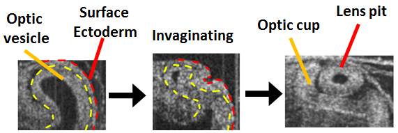
In the early embryo, the optic vesicles (OVs) emerge from the sides of the developing forebrain. As they grow, the OVs acquire an asymmetric pear shape. Eventually, they contact and stick to an exterior membrane, whereupon the adhered two layers invaginate (dimple inward). The inner layer creates the bilayered optic cup, while the outer layer closes to create the primitive lens vesicle. The optic cup eventually becomes the retina and retinal pigmented epithelium.
Abnormal eye morphogenesis can cause visual impairment associated with a number of maladies, including Peters' anomaly and retinal coloboma, sometimes leading to microphthalmia. These disorders affect significant numbers of newborns.
Using strategies like those used to study heart and brain morphogenesis, we are determining the mechanical forces that drive formation of the optic vesicle and optic cup.
|
|
3-D optical coherence tomography reconstructions show optic vesicle (OV) evagination from the forebrain (F) and subsequent eye morphogenesis in the early embryo. |
|
|
Optical Coherence Tomography images of the developing chick eye. The optic vesicle and surface ectoderm invaginate to form the optic cup and lens pit. |
|
|
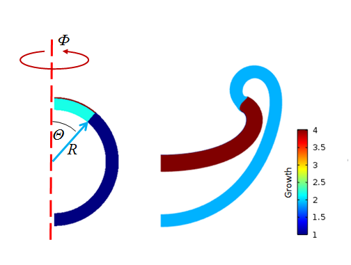
A finite element model of the eye starts as a spherical optic vesicle with a thin, stiff extracellular matrix on the top surface. The optic vesicle forms an invaginated shape as it grows. |


